Re-use of this resource is governed by a Creative Commons
Attribution-
NonCommercial-ShareAlike 4.0 International
https://creativecommons.org/licenses/by-nc-sa/4.0/
NonCommercial-ShareAlike 4.0 International
https://creativecommons.org/licenses/by-nc-sa/4.0/
Piezoelectrics are found in medicine, where they are used
in two ways:
An alternating field of a high frequency is applied to
the piezoelectric, so that its shape changing forms waves in the ultrasound
range.
Waves in the ultrasound range.
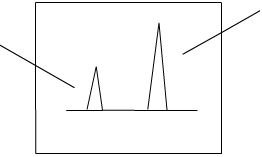
These ultrasound waves if pointed at a kidney stone can
'shake' it apart. This is a good non-invasive technique.
Ultrasound waves can reflect from tissue boundaries.
The reflected waves can be observed in order to produce an image of
internal body tissues, such as a fetus.
The piezoelectric changes shape as an applied field is
reversed. It varies with the frequency of the electric field, producing
waves in the ultrasound range.


The waves are directed onto the fetus, usually via a handheld
device.
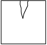
Perhaps a more relevant example of ultrasound is its use
in crack detection. The principles are the same.
The ultrasound is again produced by a piezoelectric, and
the waves reflect from both sides of possible cracks in order to detect
it.
The image is picked up by the piezoelectric, and gives an
image of the crack.
Start of crack
End of crack