Micrograph Library
Browse the libraryAdvanced searchSystemsCompositionsTechniquesKeywordsPhase diagramsHelpPreferencesAbout the micrograph libraryTerms of useContribute micrographs!FeedbackLinksCredits Print this page
Full Record for Micrograph 645
- Micrograph no
- 645
- Brief description
- An image of a single crystal alkane
- Keywords
- alkane, lamella
 , polymer , single crystal
 - Categories
- Polymer
- System
- Alkane
- Composition
- C294H590
- Standard codes
- Reaction
- Processing
- The sample was crystallized at 68 degrees C for 16 hours
- Applications
- These materials provide ideal model systems for understanding the behaviour of synthetic polymers, as they are available in lengths bridging the gap between behaviour typically associated with polymers (such as chain folding) and that of small molecules. However, in contrast to fractionated materials, they are strictly monodiperse, allowing effects of polydispersity to separated from those of chain length.
- Sample preparation
- Technique
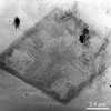
- Transmission electron microscopy (TEM)
- Length bar
- 2.4 μm
- Further information
- In high polymers, although it is commonly observed that crystalline lamellae grown from the melt thicken after initial crystallization, in crystallization from dilute solution it is generally found that the single crystals grown maintain the thickness with which they originally grew unless they are annealed above their original crystallization temperature. In contrast to this, it has been found that ultra long alkanes grown in dilute solution will thicken at the crystallization temperature from an initial (integer) folded form to more stable, thicker, integer folded or extended forms, given sufficient time. The thickening behaviour of C294H590 between three times folded and twice folded has been studied. This TEM image is of a thickened lozenge shaped crystal, clearly showing the preferential thickening of sector boundaries to form a 'picture frame' outer rim, and the dendritic structure of the central region.
- Contributor
- J Hobbs
- Organisation
- Scanning Probe Microscopy Group, Bristol University
- Date
- 03/10/02
- Licence for re-use
 DoITPoMS standard terms of use