Micrograph Library
Browse the libraryAdvanced searchSystemsCompositionsTechniquesKeywordsPhase diagramsHelpPreferencesAbout the micrograph libraryTerms of useContribute micrographs!FeedbackLinksCredits Print this page
Full Record for Micrograph 561
[91 KB]
View micrograph
.. in new window
View micrograph and record
.. in new window
You can also view and download the micrographs on Flickr
- Micrograph no
- 561
- Brief description
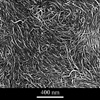
- Ordered film of carbon nanotubes
- Keywords
- alignment, carbon, carbon nanotube, dispersion, film, nanotube
 , order, polymer , suspension
 - Categories
- Ceramic, Polymer
- System
- Carbon nanotube
- Composition
- Not specified
- Standard codes
- Reaction
- Processing
- A dilute (<0.3 vol%) suspension of nanotubes is filtered through a 0.2 mm membrane to form a solid film.
- Applications
- Carbon nanotubes may be considered a high performance mechanical polymer or an electrically conducting polymer but their greatest potential is in gas storage or as a filler in polymeric materials
- Sample preparation
- Solid nanotube films were prepared by filtration onto a 0.2 mm membrane filter under 0.6 bar negative pressure
- Technique
- Field emission gun scanning electron microscopy (FEGSEM)
- Length bar
- 400 nm
- Further information
- It is necessary to form a stable dispersion of nanotubes in order to properly integrate them into polymeric systems. This can be achieved by treating them with acid to oxidise the tube surfaces. The tubes will then spontaneously disperse in an aqueous medium. The viscosity of these suspensions is analogous to that of polymers; it increases gradually with concentration up to a critical point (at about 0.7vol%) where entanglement occurs. A solid nanotube film has been formed by filtering the suspension through a 0.2mm membrane filter. Suspensions of relatively high concentration (>0.3vol%) yield films with random tube orientations but at lower concentrations (as in this sample), liquid crystal aggregation occurs and there is noticeable mutual alignment. The films exhibiting such alignment are tougher. This image was taken using a field emission gun scanning electron microscope (FEGSEM).
- Contributor
- Prof A H Windle
- Organisation
- Department of Materials Science and Metallurgy, University of Cambridge
- Date
- 03/10/02
- Licence for re-use
 Attribution-NonCommercial-ShareAlike 4.0 International