Micrograph Library
Browse the libraryAdvanced searchSystemsCompositionsTechniquesKeywordsPhase diagramsHelpPreferencesAbout the micrograph libraryTerms of useContribute micrographs!FeedbackLinksCredits Print this page
Full Record for Micrograph 563
[60 KB]
View micrograph
.. in new window
View micrograph and record
.. in new window
You can also view and download the micrographs on Flickr
- Micrograph no
- 563
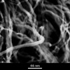
- Brief description
- Carbon nanotubes
- Keywords
- carbon, carbon nanotube, dispersion, film, nanotube
 , order, polymer , suspension
 - Categories
- Ceramic, Polymer
- System
- Carbon nanotube
- Composition
- Low concentration (<0.3 vol%) suspension in water
- Standard codes
- Reaction
- Processing
- A loose aggregate of nanotubes is treated with acids and washed. The treated tubes spontaneously disperse in water
- Applications
- Carbon nanotubes may be considered a high performance mechanical polymer or an electrically conducting polymer but their greatest potential is in gas storage or as a filler in polymeric materials
- Sample preparation
- Solid nanotube films were prepared by filtration onto a 0.2 mm membrane filter under 0.6 bar negative pressure
- Technique
- Field emission gun scanning electron microscopy (FEGSEM)
- Length bar
- 99 nm
- Further information
- It is necessary to form a stable dispersion of nanotubes in order to properly integrate them into polymeric systems. This can be achieved by treating them with acid to oxidise the tube surfaces. The tubes will then spontaneously disperse in an aqueous medium. The viscosity of these suspensions is analogous to that of polymers; it increases gradually with concentration up to a critical point (at about 0.7 vol%) where entanglement occurs. However, their separation is determined more by surface repulsions than by entropy arising from chain flexibility. Their stiffness suggests that parallel clusters might be a natural state for aggregation but it also means that any deviations form straightness of the tubes (due to defects) will compromise significant tube parallelism. This image was taken using a field emission gun scanning electron microscope (FEGSEM).
- Contributor
- Prof A H Windle
- Organisation
- Department of Materials Science and Metallurgy, University of Cambridge
- Date
- 03/10/02
- Licence for re-use
 Attribution-NonCommercial-ShareAlike 4.0 International